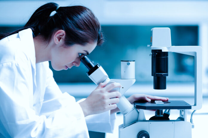
The stereo Microscope, stereoscopic or dissecting microscope is an optical microscope variant designed for low magnification observation of a sample, typically using light reflected from the surface of an object rather than transmitted through it. The instrument uses two separate optical paths with two objectives and eyepieces to provide slightly different viewing angles to the left and right eyes. This arrangement produces a three-dimensional visualization of the sample being examined.[1] Stereomicroscopy overlaps macrophotography for recording and examining solid samples with complex surface topography, where a three-dimensional view is needed for analyzing the detail.
The stereo microscope is often used to study the surfaces of solid specimens or to carry out close work such as dissection, microsurgery, watch-making, circuit board manufacture or inspection, and fracture surfaces as in fractography and forensic engineering. They are thus widely used in manufacturing industry for manufacture, inspection and quality control. Stereo microscopes are essential tools in entomology.
The stereo microscope should not be confused with a compound microscope equipped with double eyepieces and a binoviewer. In such a microscope, both eyes see the same image, with the two eyepieces serving to provide greater viewing comfort. However, the image in such a microscope is no different from that obtained with a single monocular eyepiece.
History
[edit]
The first optically feasible stereomicroscope was invented in 1892 and became commercially available in 1896, produced by Zeiss AG in Jena, Germany.[2]

American zoologist Horatio Saltonstall Greenough grew up in the elite of Boston, Massachusetts, the son of the famous sculptor Horatio Greenough Sr. Without the pressures of having to make a living, he instead pursued a career in science and relocated to France. At the marine observatory in Concarneau on the Bretton coast, led by the former director of the Muséum national d’histoire naturelle, Georges Pouchet, he was influenced by the new scientific ideals of the day, namely experimentation. While dissection of dead and prepared specimens had been the main concern for zoologists, anatomists and morphologists, during Greenough’s stay at Concarneau interest was revived in experimenting on live and developing organisms. This way scientists could study embryonic development in action rather than as a series of petrified, two-dimensional specimens. In order to yield images that would do justice to the three-dimensionality and relative size of developing invertebrate marine embryos, a new microscope was needed. While there had been attempts to build stereomicroscopes before, by for example Chérubin d’Orleans and Pieter Harting, none had been optically sophisticated. Furthermore, up until the 1880s no scientist needed a microscope with such low resolution.


